


Human Larynx Model
Human Larynx Model
A functional model that demonstrates movements of the epiglottis and cartilages in the voice box. It helps the students to require and understanding of the morphology and structure of the respiratory tract and phonetic organ. Dissectible into 3 parts, 3 times enlarged.
Size: 11.5x11x24CM.
Material: PVC


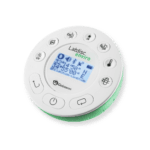 Labdisc
 Botzees
 Edison
 Telepresence Robot
 DOBOT
DOBOT